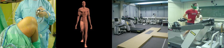
Orthopaedic Sports Medicine Center of Ioannina

“A modern and fully equipped facility”

Faqs

What is an Anterior Cruciate Ligament (ACL) rupture?

The presence of the anterior cruciate ligament (ACL) is particularly important for maintaining knee joint stability, as this ligament constitutes the fundamental factor of resistance in anterior tibial translation and internal knee rotation. ACL rupture is a very common athletic injury, and its frequency constantly increases in the last few years, as more and more people take part in sports activities.

Should we operate an ACL rupture?

In contrary to other ligaments, ACL cannot heal with simple suturing and the result of its rupture is the creation of episodes of instability and knee "giving way" during athletic or even daily activities, that require rotational knee movements. Furthermore, in an ACL deficient knee, the probability of a new meniscal injury and chondral degeneration increases, due to the pathological anterior drawer, rotatory instability and poor control of muscle function. Longitudinal follow-up studies have showed that ACL deficiency is associated eventually with post-traumatic arthritis. For this reason, surgical reconstruction of the ACL rupture is recommended in order to re-establish knee normal function. ACL reconstruction is the 6th more frequent orthopaedic surgery nowadays, in USA.

How can we diagnose an ACL rupture?

The diagnosis will be based upon the mechanism of injury (usually an abrupt rotational knee movement with the foot "stuck" in the ground), the existence of knee joint effusion, that may probably constitute the unique clinical sign, and the special tests (Lachman-Noulis, anterior-drawer, pivot-shift) that the doctor performs at the clinical examination. One special feature of the acute rupture is that the patient hears a specific noise during the injury moment, like breaking a rope and in most cases he cannot continue the athletic activity. The diagnosis is always justified with a knee MRI.

yerine gitmek Şimdi büyük kazanmak istiyorsanız Mostbet!

What is knee instability?

Knee instability describes the state in which the knee joint is at risk for subluxation. It is due to the rupture of the ligaments that stabilize the knee joint (anterior cruciate ligament, posterior cruciate ligament, collateral ligaments, and/or posterolateral complex of the knee). In these conditions, the patient feels that the knee is unstable during specific activities such as pivoting, twisting, turning, going down stairs or slopes. Treatment can be conservative or surgical depending on the kind of the injury, the overall grade of instability and the patient's activity level.

What knee arthritis mean?

Under the term knee arthritis we describe the inflammation of the knee joint. The most common cause of knee arthritis is osteoarthritis, a disease in which artcular cartilage progressively degenerates. It usually affects people of 50 - 60 years old. Posttraumatic arthritis resembles osteoarthritis. It results from trauma (fractures, ligament tears and meniscal tears). Arthritis is also encountered as a manifestation of autoimmune disorders as rheumatoid arthritis. All these conditions are characterized by absence of bacterial factor. In septic arthritis though, the cause is a bacterial infection. In sport related injuries the most common cause of arthritis is trauma. Treatment of arthritis includes conservative measures (physiotherapy, pain killers, NSAID's and glucosamine , chondroitin administration and intraarticular injection of corticosteroids and hyaluronic acid) or surgical. ( osteotomy, arthroscopic shaving, microfracture, autologous chondrocyte transplantation mosaicplasty and finally unicompartmental or total knee arthroplasty).

What is articular cartilage?

Articular cartilage covers the articulating surfaces of the bones within a joint. It is a tough, elastic tissue that enables the bones to move smoothly one over another while at the same time it absorbs a large amount of the loads applied on them. Articular cartilage lesions are due to trauma or degeneration. They range from simple fissuring to large defects in which the underlying bone is exposed. Clinical manifestations include pain, limited range of motion and in certain cases joint oedema. It has been shown that small defects can progress to larger lesions if left untreated. Treatment can be conservative (physiotherapy, pain killers, NSAID's and glucosamine administration) or surgical. (arthroscopic shaving, microfracture, autologous chondrocyte transplantation and mosaicplasty).

Which is the time interval between the injury and the first myodynamic test and what are the criteria for an athlete or sports active person to start the myodynamic control?

As far as the ligaments, muscles and tendons injuries concern, when the injured parts are involved on the function of the testing muscles, full medical tissue rehabilitation is required before the first control.
After a surgical treatment, like the ACL reconstruction, with which we mainly deal in our lab, the starting criteria are principally these:
• The surgery type. For example in the ACL reconstruction we consider that in the last knee extension degrees just as in every similar open kinetic chain exercise, increased anterior shear force is produced with a following increased danger for the graft.
• The overall rehabilitation progress. Athletes following accelerated programs are myodynamically controlled earlier than sports active people following conservative programs with limited rehabilitation sessions.
According these, the time interval for the myodynamic control beginning after an ACL reconstruction varies between 4 and 6 months in our lab.

What are the advantages from the use of ultrasound as an imaging tool?

* Ultrasound scanning is noninvasive (no needles or injections) and painless imaging modality
* Ultrasound is widely available, easy-to-use and less expensive than other imaging methods.
* Ultrasound imaging uses no ionizing radiation.
* Ultrasound scanning gives valuable information of soft tissues that cannot be visualized on plain x-ray images.
* Ultrasound causes no health problems and may be repeated as often as is necessary if medically indicated.
* Ultrasound provides real-time imaging, allowing the dynamic assessment of the examined structures and the interaction between doctor and patient.
* Unlike the strong magnetic field of magnetic resonance imaging (MRI), ultrasound is not affected by ferromagnetic implants or fragments within the body. Ultrasound is also an excellent alternative to MRI for claustrophobic patients.
* Ultrasound may actually have advantages over MRI in seeing tendon structure, which is better appreciated by ultrasound than MRI.

Exercise Testing for Athletic and Team support

Using a state of the art on-line gas analysis system, we can accurately measure VO2 and other cardiopulmonary variables during an incremental exercise test to exhaustion, in order to calculate maximal oxygen consumption (VO2 max) - a gold standard measure of aerobic fitness. During the max test, we can measure blood lactate at intervals which we use to determine training zones in order to determine short- or long-term training programs and also assess adaptation to specific training programmes through repeated testing. We can tailor a test to suit your sport, and can run a max test on treadmill or cycle ergometer. We can offer support for coaches working with teams, or other groups, and have the flexibility to perform certain field tests. Coaches may be interested in monitoring certain fitness parameters, such as maximal oxygen uptake, anaerobic capacity, muscular strength and body composition in their team players. Participants may range from professional athletes (some which are of national and international standard) to amateur athletes interested in maximizing their performance. Besides regular participation in a sport discipline, a recent medical exam is prerequisite.

Is this injury common? Does it happen more in men or in women?

It has been found that approximately one ACL rupture will occur for every 1500 player-hours spent practicing or competing in sports such as football, skiing, basketball and soccer. It has been reported that almost 250.000 ACL ruptures occur in the US every year. Female athletes are the most likely targets, with epidemiological data suggesting that females are up to six times more likely to sustain such an injury. Thus, ACL rupture is a common sports-related injury, especially in female athletes.